Are you eyeing a new site for optical care?
MU staff and faculty have a new eye care facility to visit with more space and state-of-the-art eye equipment. University Eye Institute East opened Jan. 9 at 3215 Wingate Court near MU’s Women’s and Children’s Hospital.
The institute significantly expands the eye care services that were offered at the old facility, University Physicians Eye Institute East, located on Portland Ave. on the west side of the hospital. The 7,900-square-foot center is triple the size of the old building.
Sue Mussatt, administrative associate at MU’s Mason Eye Institute, worked with a committee to design the building. Mussatt and the committee planned several child-friendly areas in the institute. The waiting room in the new facility includes a separate play area where children can watch TV or play with toys.
A special section of the institute, separate from the adult examination rooms, is dedicated to children’s eye care. The section includes six rooms, each having a unique decorative design.
Pictures of TJ, the Children’s Hospital tiger mascot, getting an eye exam will soon adorn the walls. Gaye Baker, reimbursement coordinator for MU’s Department of Ophthalmology, said the idea is to reduce children’s fear.
“We’ll do anything that helps reduce kids’ anxiety when they’re going through something that they believe to be very stressful,” Baker said.
Adjacent to the waiting room is University Optical East, where patients can purchase prescription frames, sunglasses, pediatric glasses, contact lens and other eye-care products.
The additional space allows for a staff up to 20. Four attending physicians work among four exam rooms during a shift. Working toward their medical degrees, residents and fellows assist the attending physicians. A cornea specialist and physicians technicians, who conduct lab tests, also are on staff.
New Technology
The new facility also has several state-of-the-art pieces of technology.
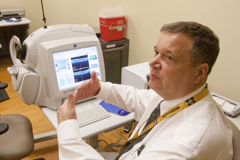
One is the Fundus camera, which takes photos of the retina and other parts of the eye. Coy Cobb, facilities coordinator, uses the machine to detect epiretinal membrane, also known as macular pucker, a scar tissue that covers the macula and causes blurred vision.
Another piece is the optimal coherence tomography (OCT) machine, which maps the back of the eyes.
“If you think of geology and that you’re doing core samples and you’re looking at various levels in the soil, this looks at the levels at the back of your eye,” Baker said.
The patient looks into a lens on the machine and fixates on a crosshair. The light bounces off the back of the eye and back into the machine where it is read. Each layer of the eye reflects the light differently. Cobb then analyzes the layers down to the micron level.
Before the technology’s development in 1992, this type of examination was impossible, Cobb said.
“This is the latest and greatest version of the OCT,” he said. “There’s no better in the entire country than what you see right here.”
The OCT can detect glaucoma, which Baker calls “the silent stealer of sight” because it shows few symptoms. In the facility’s two visual field rooms, physicians can detect visual impairment when patients cannot.
Eye exams are recommended every two years, Mussatt said.
— Trevor Eischen